High hamstring tendinopathy: a pain in the bum

The last installment in our series on tendinopathy takes a look at the proximal hamstring tendon. A growing number of running athletes suffer from the degenerative tendon disease known as proximal hamstring or high hamstring tendinopathy (HHT)(1) . Athletes complain of a pain at the gluteal fold, which may not bother them during running but ache afterwards, especially during long periods of sitting. They may not know of a precipitating incident but a careful history usually reveals a sudden increase in training load or change in training environments, such as integration of hill training or track sprints. Physical exam may also reveal an overly powerful quadriceps muscle, leaving the hamstring straining to compensate for the imbalance.
As writer and sports therapist, Trevor Langford, reviews here, the hamstring consists of the semimembranosus, semitendinosus, and the long head of the biceps femoris, all of which originate from the ischial tuberosity, and the short head of the biceps femoris which originates from the femur. The hamstrings insert on either side of the knee on both the tibia and the fibula. A positive ‘take the shoe off’ test is highly specific for hamstring injuries and often reproduces the pain at the ischial tuberosity (see figure 1).
Figure 1: Shoe off test
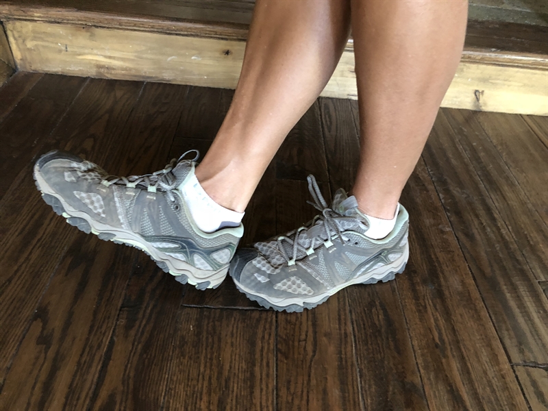
When an athlete tries to remove a shoe pulling back against the other foot, pain in the hamstring denotes a positive 'shoe off' test.
Treatment
Langford illustrates a typical progression of conservative management of an athlete with HHT, emphasizing progressive eccentric loading. One challenge in treating these injuries is managing hamstring load in the athlete’s daily activities. Hip flexion places a stretch on the tendon, and thus loads the site. Athlete’s in the acute stage should avoid sitting for long periods of time. Suggest standing at a desk, frequent walk breaks, or reclining chairs to decrease hip flexion. Athletes who receive conservative management follow the typical rehabilitation course of 12 weeks to return to pain free function. Again, education and managing expectations are of the highest priority to ensure safe return to play and avoid repeated injury.Shockwave therapy, the same treatment initially developed for the treatment of kidney stones, is another tool used to treat HHT. An Italian study published in 2011 but conducted in 2006, found that athletes with HHT who received shockwave therapy fared better than those randomly assigned to receive conservative therapy(2). While positive outcomes came from the shockwave branch of the study, a look at the conservative management reveals that the treatment that group received is outdated. Therefore, while the adverse effects of shockwave therapy are minimal, the evidence for its use over conservative management is lacking. Furthermore, in the United States, medical insurance doesn’t typically cover its use for this injury(3).
Lastly, athletes may resort to injection therapy to manage either the acute pain, or the chronic nature of HHT. As Langford points out, while initially effective, conservative management using heavy slow resistance training showed more positive results in the long term. Injection therapy is not without risk, as Tracy Ward explores here (see table 1). Therefore, it should be undertaken judiciously in athletes who simply can not tolerate movement or manage the pain.
Table 1: Side effects and risks of injection therapy
| Side effects and risks |
|---|
| All invasive procedures carry side effects and risks. However if the athlete is screened correctly and appropriately selected for the injection then the risks are low. Below is a summary of the main issues to consider: |
| Side effects: -Bleeding/bruising/pain at the injection site and surrounding area;-Sepsis — figures report a 1 in 17 to 1 in 162,000 chance of developing local infection(5); -Anaphylaxis — severe allergic reactions are very rare but caution should still be exercised(6);-Skin depigmentation — skin colour changes and atrophy can occur in approximately 4% of cases but are rarely significant(7). |
| Risks:-Corticosteroids are toxic to the cells that form cartilage and collagen, and their injection will inhibit collagen formation and cause death of these cells. This process can occur for the first 7 days post-injection(8). During this time the tissue rupture risk is increased. Healthy cartilage should never be injected as it may be detrimental to the healing process; therefore an accurate diagnosis is required promptly;-If an injection fails to improve symptoms a second injection may be performed, but usually a maximum limit of three injections within one year is recommended. This may be because of these risks and on-going disturbance to the tissues, but also because it may suggest the injection is not the answer to recovery and additional rehabilitation methods should be explored to identify the root of the problem. |
SIB Final word
As we close out this series, an overwhelming theme runs through. Tendinopathy, once taken hold, requires time and loading to heal. Other therapies, such as NSAIDs and injections can be deployed to help manage the pain, but they don't hurry the healing process. All patients fare better when they are educated and know what to expect. Therefore, don't make false promises or fall into the trap of thinking some magical treatment will accelerate healing. The best way to treat tendinopathy is to avoid tendon strain in the first place by making smart training decisions. However, when it can't be avoided, progressive loading through eccentric contractions or heavy slow resistance training over a 12 week period is the best strategy to safely return an athlete to their previous level of play, no matter which tendon is injured.References
- Br J Hosp Med (Lond). 2018 Jul 2;79(7):389-394.
- Am J Sports Med 2011;39:146-153.
- PM R. 2018 May 31. pii: S1934-1482(18)30237-5 [Epub ahead of print]
You need to be logged in to continue reading.
Please register for limited access or take a 30-day risk-free trial of Sports Injury Bulletin to experience the full benefits of a subscription. TAKE A RISK-FREE TRIAL

Alicia Filley

Latest Issue

Subscribe Today
Newsletter Sign Up
Subscriber Testimonials
Dr. Alexandra Fandetti-Robin, Back & Body Chiropractic
Elspeth Cowell MSCh DpodM SRCh HCPC reg
William Hunter, Nuffield Health
Subscribe Today


Newsletter Sign Up


Coaches Testimonials
Dr. Alexandra Fandetti-Robin, Back & Body Chiropractic
Elspeth Cowell MSCh DpodM SRCh HCPC reg
William Hunter, Nuffield Health
Subscribe Today
Latest Issue
Be at the leading edge of sports injury management
Our international team of qualified experts (see above) spend hours poring over scores of technical journals and medical papers that even the most interested professionals don't have time to read.
For 17 years, we've helped hard-working physiotherapists and sports professionals like you, overwhelmed by the vast amount of new research, bring science to their treatment. Sports Injury Bulletin is the ideal resource for practitioners too busy to cull through all the monthly journals to find meaningful and applicable studies.
*includes 3 coaching manuals
Get Inspired
All the latest techniques and approaches
Sports Injury Bulletin brings together a worldwide panel of experts – including physiotherapists, doctors, researchers and sports scientists. Together we deliver everything you need to help your clients avoid – or recover as quickly as possible from – injuries.
We strip away the scientific jargon and deliver you easy-to-follow training exercises, nutrition tips, psychological strategies and recovery programmes and exercises in plain English.