
 close
close


 LOGIN
LOGINAdductor Squeeze Test: Don’t Miss the Point
The adductor squeeze test sits on almost every preseason screen and in-season monitoring sheet in team sports. It is quick, cheap, and reliable. But a low score is a question, not a diagnosis. Eurico Marques sets out a clinical reasoning framework for working out why a squeeze is low and why the reflex of reaching straight for adductor strengthening leaves so many groins unresolved.



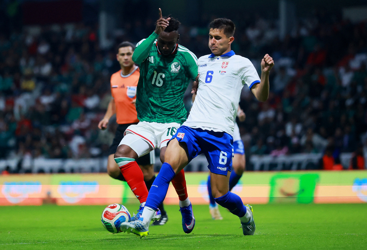
Club Leon midfielder Juan Dominguez (8) kicks the ball in the second half against the Real Salt Lake during Leagues Cup Quarterfinals at Dick’s Sporting Goods Park. Mandatory Credit: Ron Chenoy-Imagn Image
Groin pain is one of the most stubborn problems in field and court sport(1). Over a single season, close to half of male footballers report hip or groin pain, and around one in three carry symptoms into the following season(2). Small wonder that the adductor squeeze test has become a fixture of screening and monitoring across football, rugby, ice hockey, Gaelic and Australian Rules football(3-7).
The typical decision process runs like this: test, find a low score, prescribe adductor strengthening. That logic rests on two assumptions: that a low squeeze means weak adductors, and that stronger adductors will fix the problem. However, both deserve challenging. A recent framework paper from performance and clinical specialists working in elite football and at a high-volume orthopedic and sports medicine hospital in Doha argues that this reflex reflects a poor understanding of what actually drives a squeeze score(1). When clinicians target a single contributor to a multifactorial outcome, the results are unpredictable.
“Match the exercise to the deficit...”
What the Squeeze Actually Measures
The squeeze test measures the total force an athlete can produce in the frontal plane by pressing both legs together against a resistance in a set position (see figure 1). Crucially, that force is not the adductors alone. The adductor group includes pectineus, adductor longus, gracilis, adductor brevis, and adductor magnus, with secondary contributions from the long head of biceps femoris, the posterior fibers of gluteus maximus, quadratus femoris, and obturator externus(8). Worth noting is that there is also a likely contribution from the ankle and hamstrings during long-lever testing. Beyond the muscles themselves, the force registered depends on trunk control, pelvic position and the athlete’s ability to coordinate force production across segments(1,9). A low score can originate anywhere along that chain, which is exactly why it cannot, on its own, tell you where the problem sits.
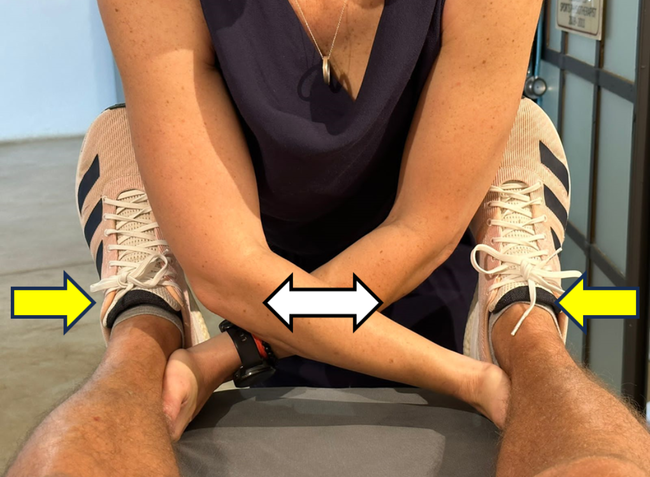
Force and Pain
In an athlete with pain, the squeeze produces two outcomes: force and pain. A gain in squeeze function can therefore show up in three ways: the same pain but more force, the same force but less pain, or progress on both fronts at once(1,10). Therefore, treating the squeeze as a single number throws away that information. Clinicians must record both force and pain on every squeeze test. They are separate variables that tell you separate things.
Testing Reasoning
Before deciding what a low score means, clinicians must question why they are testing at all. The squeeze test serves three distinct purposes, and each produces a different kind of “low score” (see table 1).
- Diagnose - As a diagnostic or early-warning test, it is a sensitive pain-provocation test for the adductor–pubic region. So, it will usually pick up a problem in these tissues, but it is not specific; i.e., pain on a squeeze will not reliably tell you which structure is responsible(11). A pain-free squeeze, by contrast, helps rule out the region.
- Screen - As a screening tool, it gives a baseline reference value: force below 465 N (or 7.0 N/kg) is associated with a substantially higher probability of groin injury in elite football, and an adductor-to-abductor ratio below 0.80 with a 17-fold increase in adductor strain risk in professional ice hockey(3,5). Large normative databases now provide percentile reference points by sex, and sport-specific preseason data provide further context(12,13).
- Monitor - As a monitoring tool, serial testing across the season tracks an athlete’s response to training and match load, where a drop from a known baseline is the signal that something has changed(14).
Table 1: The three purposes of the squeeze test.
| Purpose | What it gives the clinician | The kind of "low score" |
| Diagnositc/early warning | Pain provocation in the adductor–pubic region; sensitive but not specific | A painful squeeze that flags regional irritability |
| Screening (baseline) | A reference value versus norms and the squad | A below-benchmark score at first test |
| Monitoring (in-season) | Response to training and match load over time | A drop from the athlete’s own baseline |
Three things the squeeze cannot tell clinicians
- It is bilateral. The test yields a single combined number and is blind to side-to-side deficits, which are common and clinically relevant.
- It is isometric and tested in inner range. Force is captured statically, typically at 45° of hip flexion(15). Yet, most adductor injuries occur during outer-range adduction combined with external rotation, either while kicking or on the stance leg when changing direction(16). Eccentric strength discriminates symptomatic players better than isometric strength(17). For example, no clinician would assess a hamstring solely in deep knee flexion and then pronounce on how it behaves at long muscle lengths.
- It ignores lever-arm length. A 200cm AFL player and a 176cm footballer producing the same force are not producing the same joint moment. Absolute and even body-mass-normalized thresholds can mislead across statures.
Low Score Analysis
Step back from the complexity, and there are fundamentally four reasons an athlete’s squeeze can be low(1).
- Pain: the athlete has pain during the test, and inhibition limits their output. This is a pain problem with reduced force, not necessarily a deficit in the capacity of the adductors themselves.
- A direct deficit: the adductors genuinely lack the capacity to produce force, whether through structural factors (atrophy, tissue quality) or reduced neural drive.
- An indirect deficit: something other than adductor strength is limiting the system. For example, the stabilizers that enable force production (obliques, gluteus minimus, deep hip rotators), the synergists that contribute (psoas, gluteus medius, hamstrings, gluteus maximus), or the central nervous system’s capacity to coordinate the action (fatigue, post-match central depression, chronic deconditioning).
- Test execution: the athlete simply did not produce a true maximal effort, through poor cueing, unfamiliarity with the test, or an inadequate warm-up.
Clinicians should rule out test execution first, as a squeeze only has value if the effort is maximal. After that, the distinction between direct and indirect is the one that matters, because it determines the intervention. That distinction can only be made through a structured assessment, never from the squeeze number itself.
“A deficit in a small stabilizer can limit the output of a much larger prime mover.”
The Evidence: Strength Explains Surprisingly Little
Two lines of evidence undercut the “weak squeeze = strengthen adductors” logic. The first is biomechanical. The actions of the hip muscles vary with the position of the pelvis relative to the femur, and altering pelvic tilt significantly affects hip torque production and agonist–antagonist force ratios, even in healthy individuals performing the same test(9,18). In other words, two athletes with identical adductor strength can produce different squeeze scores purely because of differences in pelvic control.
Small stabilizing muscles also coordinate the environment in which larger muscles operate: quadratus femoris produces little absolute force yet is highly active during walking and running, stabilizing the femoral head, and hamstring EMG rises substantially as running speed increases(19,20). A deficit in a small stabilizer can limit the output of a much larger prime mover.
The second line of evidence is clinical, and it comes from injured athletes. When rehabilitation targets intersegmental control rather than the adductors directly, squeeze scores still improve. In one study of 42 athletes with athletic groin pain and 36 matched controls, every baseline hip-strength deficit resolved, yet the change in hip strength explained only 11% of the improvement in patient-reported outcomes, and the change in adductor strength was not even retained by the statistical model(21). A larger series of 205 consecutive patients rehabilitated on the same principles saw squeeze values rise and 89% return to play pain-free by 10 weeks(22). This points to a conceptual switch: when a squeeze improves after this kind of work, it need not mean the adductors grew stronger; it means the system became better at producing force in the test position(1).
The Decision Tree
The full framework is best followed using a flowchart (see figure 2)(1). When the squeeze is painful, the immediate question is diagnosis: what is the source of the pain, and does the athlete need medical or rehabilitative management? Ongoing squeeze testing, force, and pain together then track rehab progress and support transition decisions and return to play. As symptoms settle, the painful pathway feeds into the same full assessment used for pain-free presentations.
When the squeeze is low but pain-free, context decides the next move. If the score has dropped from a known baseline (monitoring context), remember that post-match reductions typically recover within 48–72 hours(14). Recovery comes first: modify participation, then re-test. If the score improves, continue the program; if it remains low despite load adjustments, the athlete enters the full assessment.
If, instead, the score was low on the first test (in a screening context), the question is where the athlete sits relative to the expected norm. Reference data suggest a starting point (below 465 N or 7.0 N/kg; an adductor-to-abductor ratio below 0.80)(3,5). Clinicians should react to confirmed trends, not isolated readings: a change only counts as real once it exceeds roughly twice the test’s typical measurement error(23). In addition, players with longstanding symptoms show meaningfully lower preseason strength that relates more to symptom duration than to injury history itself(13,24).
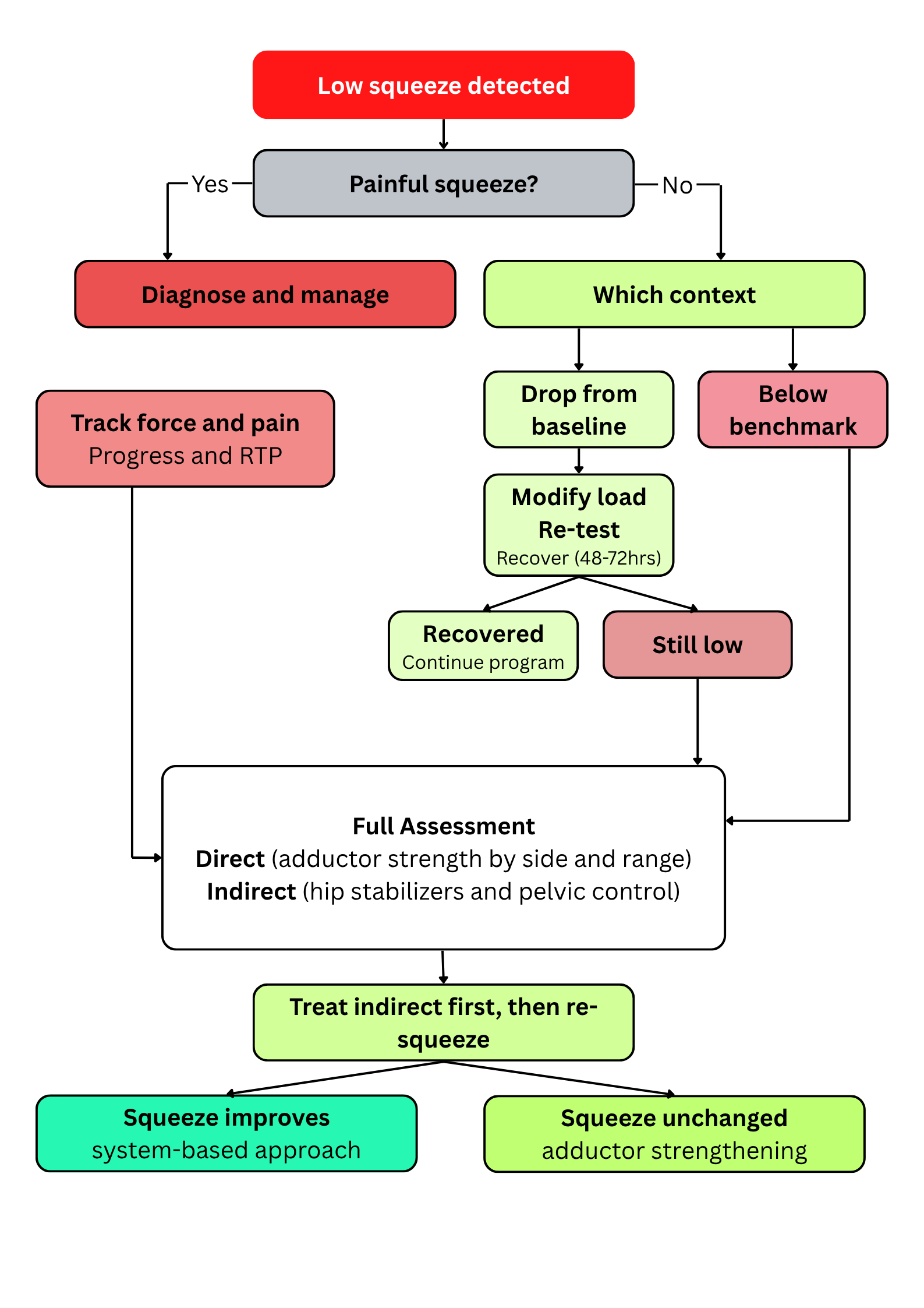
Direct and indirect deficits are presented as separate pathways for clarity, but athletes rarely fall cleanly into one category. An athlete with a genuine adductor deficit will almost always have developed compensatory coordination patterns around it, and one whose primary limitation is indirect will often show some adductor deconditioning as a consequence. The decision tree identifies the dominant limiting factor that clinicians can prioritize, but both components usually need addressing.
“When clinicians target a single contributor to a multifactorial outcome, the results are unpredictable.”
Intervention
Two principles should guide intervention, regardless of the specific exercises the clinicians choose (see table 2)(1). When addressing indirect contributors, the exercise selection should immediately change or resolve the indirect test finding. If it does not, either the exercise was wrong or the way it was coached did not achieve the intended effect. Only once the indirect test has changed can clinicians re-squeeze and read its contribution to the overall score.
In a clinical setting, this can be built into the assessment itself: test the squeeze, identify and address the indirect deficit, confirm the indirect test has changed, then re-squeeze before the athlete leaves the room. These within-session changes are acute neuromuscular responses; they need progressing and periodizing over time to become permanent.
When addressing direct adductor deficits, the exercise should be pain-free, the sensation should be felt in the targeted muscle group, and the range and contraction mode should match the deficit identified. For example, performing supine ball squeezes will not build outer-range or sagittal-plane adductor strength. Match the exercise to the deficit: which leg, which range, which contraction mode. It matters more that the athlete achieves consistent, measurable change than selecting any one special exercise.
Table 2: Likely interpretations and clinical actions for common squeeze presentations
| Finding | Likely interpretation | Do | Don’t |
| Painful squeeze, low force | Pain-limited output | Diagnose source, manage irritability, track force and pain | Chase force into pain |
| Pain-free drop post-match | Fatigue/recovery | Modify load, re-test at 48–72h | Label it adductor weakness |
| Pain-free persistent low score | Direct or indirect deficit | Full assessment | Default to adductor work |
| Squeeze improves after indirect work | System contributor likely | Progress intersegmental control | Assume adductors were weak |
| Squeeze unchanged after indirect work | Direct deficit more likely | Adductor loading by side/range/mode | Rely on short-lever ball squeezes |
Clinical Take-aways
Clinicians can implement three changes that take five minutes. They can:
- Record both force and pain on every squeeze test.
- When a score is low, ask “painful or painless?” before choosing an intervention.
- Before prescribing adductor strengthening, check at least one indirect contributor.
Adductor strengthening is the right answer when a direct deficit is confirmed, but never in isolation; always alongside intersegmental control work. Clinicians must be more critical of a low squeeze score, and defaulting to adductor strengthening may be the inappropriate reflex.
References
1. Sport Perform Sci Rep. 2026;302:v1.
2. Scand J Med Sci Sports. 2017;27(1):107-114.
3. Phys Ther Sport. 2019;37:54-59.
4. J Strength Cond Res. 2016;30(2):344-350.
5. Am J Sports Med. 2001;29(2):124-128.
6. J Sci Med Sport. 2014;17(2):155-159.
7. J Sci Med Sport. 2016;19(9):766-770.
8. J Orthop Sports Phys Ther. 2010;40(2):82-94.
9. Phys Ther. 1986;66(3):351-359.
10. J Sci Med Sport. 2021;24(11):1123-1129.
11. Br J Sports Med. 2016;50(7):423-430.
12. VALD. Practitioner’s Guide to Preseason. Volume 1. 2024.
13. Orthop J Sports Med. 2018;6(1):2325967117747275.
14. Sport Perform Sci Rep. 2017;7(1).
15. Man Ther. 2011;16(5):476-480.
16. Br J Sports Med. 2019;53(3):158-164.
17. Orthop J Sports Med. 2014;2(2):2325967114521778.
18. J Funct Morphol Kinesiol. 2024;9(2):73.
19. J Biomech. 2015;48(12):3433-3439.
20. J Sports Sci Med. 2024;23(3):603-610.
21. Am J Sports Med. 2021;49(11):2994-3003.
22. Br J Sports Med. 2018;52(16):1054-1062.
23. Sports Med. 2000;30(1):1-15.
24. J Sci Med Sport. 2017;20(4):339-343.


Latest Issue

Subscribe Today
Newsletter Sign Up
Subscriber Testimonials
Dr. Alexandra Fandetti-Robin, Back & Body Chiropractic
Elspeth Cowell MSCh DpodM SRCh HCPC reg
William Hunter, Nuffield Health
Subscribe Today

Newsletter Sign Up
Coaches Testimonials
Dr. Alexandra Fandetti-Robin, Back & Body Chiropractic
Elspeth Cowell MSCh DpodM SRCh HCPC reg
William Hunter, Nuffield Health
Subscribe Today
Latest Issue

Be at the leading edge of sports injury management
Our international team of qualified experts (see above) spend hours poring over scores of technical journals and medical papers that even the most interested professionals don't have time to read.
For 17 years, we've helped hard-working physiotherapists and sports professionals like you, overwhelmed by the vast amount of new research, bring science to their treatment. Sports Injury Bulletin is the ideal resource for practitioners too busy to cull through all the monthly journals to find meaningful and applicable studies.
*includes 3 coaching manuals
Get Inspired
All the latest techniques and approaches


Sports Injury Bulletin brings together a worldwide panel of experts – including physiotherapists, doctors, researchers and sports scientists. Together we deliver everything you need to help your clients avoid – or recover as quickly as possible from – injuries.
We strip away the scientific jargon and deliver you easy-to-follow training exercises, nutrition tips, psychological strategies and recovery programmes and exercises in plain English.
