
 close
close


 LOGIN
LOGINYou are viewing 1 of your 1 free articles
The Foundation: Skeletal Muscle Physiology
Samantha Nupen explores the current understanding of skeletal muscle physiology and its clinical relevance for practitioners working in sports medicine and rehabilitation sciences.



Melissa Jefferson-Wooden of the U.S., Jamaica’s Tina Clayton and Poland’s Ewa Swoboda in action during the Women’s 100m REUTERS/Aleksandra Szmigiel
A clear understanding of skeletal muscle physiology forms the foundation for the treatment, management, and rehabilitation of athletes and physically active individuals. Advances in muscle biology have significantly expanded classical models of muscle function, particularly in areas such as calcium regulation, fiber-type plasticity, and fatigue mechanisms.
While traditional models of skeletal muscle physiology focused primarily on sarcomeric organization and excitation–contraction coupling (ECC), emerging research has broadened our understanding of calcium handling, fiber plasticity, and fatigue mechanisms(1,2). This review integrates foundational principles of skeletal muscle structure and function with contemporary advances in calcium entry pathways, including store-operated calcium entry (SOCE) and excitation-coupled calcium entry (ECCE).
Introduction
Skeletal muscle accounts for approximately 40% of total body mass and represents 50–75% of total body protein content(3–5). Beyond enabling voluntary movement, skeletal muscle plays a crucial role in systemic metabolism, including glucose regulation, thermogenesis, and endocrine signaling(3).
Understanding the structural and physiological properties of skeletal muscle provides essential insights for clinicians involved in performance training, injury rehabilitation, and neuromuscular disease management.
Structural Organization
Skeletal muscle displays a hierarchical structure that extends from the whole muscle to the molecular components of the sarcomere(3,4). Each muscle is composed of bundles of muscle fibers, which themselves contain myofibrils, which are composed of repeating contractile units known as sarcomeres.
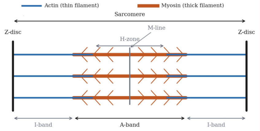
The sarcomere, bounded by Z-discs, is the fundamental contractile unit of skeletal muscle. Within this structure, thick filaments composed of myosin interact with thin filaments composed primarily of actin, along with regulatory proteins such as tropomyosin and the troponin complex (see figure 1)(3,4).
“A clear understanding of skeletal muscle physiology forms the foundation...”
These contractile elements are supported by surrounding connective tissue structures, often referred to collectively as myofascia, which provide mechanical scaffolding and contribute to force transmission within the muscle.
The Sliding Filament Mechanism
The mechanism of muscle contraction was first described in 1954 by two independent research groups working at MIT and Cambridge University(6). Their work led to the formulation of the sliding filament theory, which remains central to modern muscle physiology.
The theory proposes that during contraction, the thick (myosin) and thin (actin) filaments slide past each other rather than shortening themselves. As this occurs, the I-band and H-zone decrease in length while the A-band remains constant.
Researchers at Cambridge later expanded this concept with the cross-bridge model, proposing that myosin heads cyclically bind to actin, generate force through an ATP-driven power stroke, and then detach to repeat the cycle(6). Electron microscopy later confirmed the presence of overlapping thick and thin filaments within the sarcomere, providing structural support for this theory.
The Length–Tension Relationship
The relationship between sarcomere length and force generation is a fundamental principle of muscle physiology. Researchers at University College London demonstrated this relationship in 1966 using isolated frog muscle fibers(7).
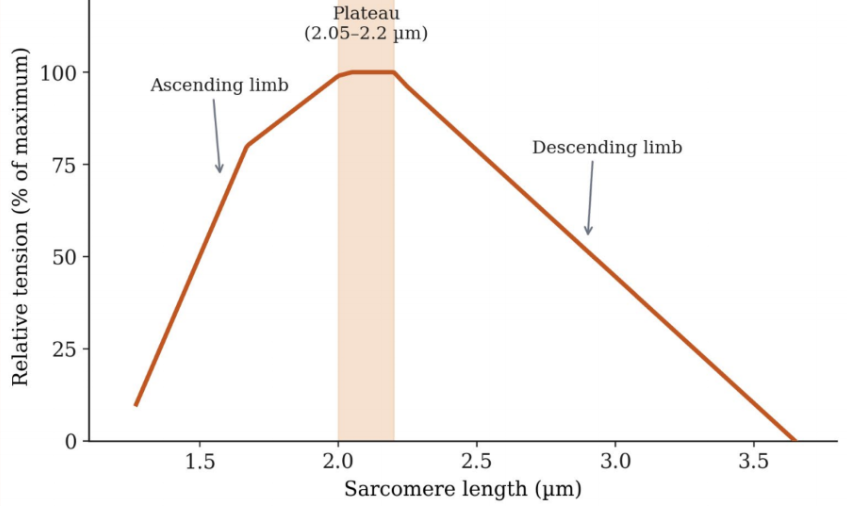
Their experiments showed that the force generated by a muscle fiber depends directly on the degree of overlap between actin and myosin filaments(8). Three key regions define the length–tension curve (see figure 2):
- Plateau region (≈2.05–2.2 µm): Maximum tension occurs when optimal overlap between actin and myosin allows maximal cross-bridge formation.
- Descending limb (>2.2 µm): Force decreases as sarcomere length increases and filament overlap diminishes.
- Ascending limb (<2.05 µm): Force decreases as excessive filament overlap interferes with effective cross-bridge formation.
“...optimal strengthening typically occurs when muscles operate near the plateau region...”
Passive tension, generated largely by non-contractile structures such as connective tissue and structural proteins like titin, contributes increasingly to total force at longer muscle lengths.
Clinically, the length–tension relationship explains why muscles produce less force when excessively shortened or excessively stretched. From a rehabilitation perspective, optimal strengthening typically occurs when muscles operate near the plateau region of the force–length curve.
Sarcomere
Subsequent research has refined classical interpretations of the length–tension relationship. One important factor is sarcomere non-uniformity, where individual sarcomeres within a muscle fiber do not maintain identical lengths during contraction. In such conditions, stronger sarcomeres may shorten while weaker ones stretch, producing a phenomenon known as ‘creep’, or a slow rise in tension during isometric contraction. This interaction allows the fiber to sustain greater force at longer lengths than simple overlap theory predicts.
Another important factor is lattice spacing, the radial distance between actin and myosin filaments. Because muscle fibers maintain approximately constant volume, changes in sarcomere length alter lattice spacing. When the sarcomere shortens, the muscle fiber expands radially, increasing lattice spacing and reducing the probability of cross-bridge formation. Conversely, when the sarcomere lengthens toward optimal length, lattice spacing decreases, facilitating cross-bridge interaction and enhancing force production.
Changes in lattice spacing may account for 20–50% of the variation in force observed along the ascending limb of the force–length curve. Additionally, myofilament calcium sensitivity is length-dependent, with greater sensitivity at longer sarcomere lengths. This further contributes to the steep rise in force generation as muscles approach their optimal length.
Excitation–contraction Coupling
Excitation–contraction coupling (ECC) is the physiological process that links electrical stimulation of a muscle fiber to mechanical contraction(3,4). The process begins when an action potential travels along a motor neuron and reaches the neuromuscular junction. The release of acetylcholine depolarizes the muscle fiber membrane (sarcolemma), generating an action potential.
To ensure rapid activation of the entire muscle fiber, the electrical signal propagates deep into the fiber via the transverse tubules (T-tubules)(9). Voltage changes in the T-tubules activate dihydropyridine receptors (DHPRs), which function as voltage sensors in skeletal muscle. These receptors are mechanically coupled to ryanodine receptor type-1 (RYR1) calcium channels located in the sarcoplasmic reticulum.
The conformational change in DHPR triggers the opening of RYR1 channels, resulting in rapid release of calcium ions (Ca2+) from the sarcoplasmic reticulum into the cytosol.
Calcium binds to troponin C, initiating a conformational change that shifts tropomyosin away from actin binding sites. This allows myosin heads to attach to actin, initiating cross-bridge cycling and force generation(9). Relaxation occurs when calcium is actively transported back into the sarcoplasmic reticulum by the SERCA (sarco/endoplasmic reticulum calcium ATPase) pump(3).
Cross-bridge Cycling and Force Generation
Force generation during contraction is driven by the cyclical interaction between actin and myosin filaments. Each cross-bridge cycle involves four key stages:
- ATP binding,
- ATP hydrolysis,
- Power stroke,
- Detachment(6).
The overall force generated by a muscle depends on both motor unit recruitment and firing frequency (rate coding). The nervous system generally follows Henneman’s size principle, recruiting smaller motor units before progressively activating larger, high-threshold units as force demands increase(3).
Different contraction types involve distinct recruitment strategies:
- Concentric contraction: Muscle shortens while generating force. These contractions typically require the greatest motor unit recruitment and firing rates for a given load.
- Eccentric contraction: Muscle lengthens while under tension and can produce 20–40% greater force than concentric contractions. This increased force partly reflects contributions from passive structural components such as titin. Eccentric contractions are also metabolically efficient and require relatively lower neural activation to generate high force.
- Isometric contraction: The muscle generates tension without changing length. Recruitment patterns typically fall between those observed in concentric and eccentric contractions.
Muscle Fiber Types
Skeletal muscle fibers are classified according to their contractile and metabolic characteristics(10). Type I fibers are slow-twitch, oxidative fibers with high fatigue resistance and high mitochondrial density. Type IIa fibers are fast oxidative-glycolytic fibers with both aerobic and anaerobic capacity. Finally, type IIx fibers are fast glycolytic fibers capable of generating high force but prone to rapid fatigue(10). Fiber type distribution is partly genetically determined but remains highly adaptable to training.
Training-induced Muscle Adaptations
Skeletal muscle demonstrates remarkable plasticity, allowing fibers to adapt metabolically and structurally in response to training stimuli. Endurance training promotes a shift toward oxidative metabolism. This includes increased mitochondrial density, greater capillary supply, and elevated oxidative enzyme activity(11). Fast-twitch fibers may transition toward a more fatigue-resistant Type IIa phenotype.
Resistance training primarily induces muscle hypertrophy, particularly within Type II fibers. This adaptation increases cross-sectional area and enhances maximal force production. Hybrid fiber types may also emerge during training, reflecting transitional states between fiber categories. Detraining reverses many of these adaptations, with fibers gradually returning toward their baseline phenotype.
Energy Systems
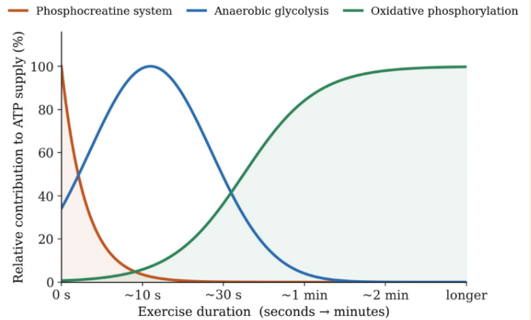
Muscle contraction requires continuous ATP availability(3). Immediate ATP stores within the muscle are limited and are rapidly replenished through three major energy systems (see figure 3):
● Phosphocreatine system provides rapid ATP regeneration for short, high-intensity activity.
● Anaerobic glycolysis generates ATP without oxygen during moderate-duration activity.
● Oxidative phosphorylation supports prolonged activity through aerobic metabolism(3,11).
“Skeletal muscle demonstrates remarkable plasticity...”
Muscle Fatigue
Muscle fatigue is defined as a reversible reduction in force-generating capacity during sustained activity(1). Understanding these mechanisms is particularly relevant for clinicians designing rehabilitation programs or managing high-performance athletes. Multiple mechanisms contribute to fatigue, including:
- Reduced calcium release from the sarcoplasmic reticulum(1),
- Accumulation of inorganic phosphate within muscle fibers(12),
- Alterations in cross-bridge cycling kinetics(13),
- Impaired membrane excitability(14).
Emerging Insights in Calcium Regulation
Traditional models of muscle physiology emphasize calcium release exclusively from the sarcoplasmic reticulum. However, recent research has highlighted the importance of extracellular calcium entry pathways.
Two key mechanisms have been identified:
- Store-operated calcium entry (SOCE): SOCE is activated when calcium stores within the sarcoplasmic reticulum become depleted. The protein STIM1 senses this depletion and activates ORAI1 channels in the cell membrane, allowing extracellular calcium to enter the muscle fiber(2,15,16). Furthermore, SOCE plays an important role in maintaining calcium homeostasis during sustained muscle activity and is essential for normal muscle development and contractile function(16,17).
- Excitation-coupled calcium entry (ECCE): ECCE represents another pathway for calcium influx during repetitive stimulation, further contributing to calcium regulation during prolonged activity(15).
Recent studies examining calcium handling within the T-tubular system have also demonstrated fiber-type differences in calcium regulation between fast- and slow-twitch fibers(18).
Clinical Implications
Understanding skeletal muscle physiology has direct relevance for clinical practice. Knowledge of fiber-type distribution, recruitment patterns, and metabolic pathways informs exercise prescription and training design(11). Insights into excitation–contraction coupling and calcium regulation are also relevant to the diagnosis and management of neuromuscular disorders, including conditions associated with mutations in the RYR1 gene, which can increase susceptibility to malignant hyperthermia during anesthesia(19).
For clinicians working in sports rehabilitation, fiber-type composition and training adaptations influence fatigue resistance, recovery, and injury risk across different athletic populations.
“Detraining reverses many of these adaptations...”
Conclusion
Skeletal muscle function depends on the precise integration of structural organization, excitation–contraction coupling, and tightly regulated calcium signaling pathways(3–5,9).
Advances in calcium physiology, particularly the discovery of extracellular calcium entry pathways such as SOCE and ECCE, have expanded classical models of muscle function and provided new insights into fatigue, training adaptation, and neuromuscular disease(2,15–17).
For clinicians working in sports medicine and rehabilitation, a strong understanding of these physiological mechanisms provides an essential foundation for effective exercise prescription, injury management, and performance optimization.
References
1. Physiol Rev. 2008;88(1):287–332.
2. Physiol Rev. 2015;95(4):1383–436.
3. Guyton and Hall Textbook of Medical Physiology. 14th ed. Elsevier; 2021.
4. Medical Physiology. 3rd ed. Elsevier; 2017.
5. Vander’s Human Physiology. 15th ed. McGraw-Hill; 2019.
6. Prog Biophys Biophys Chem. 1957;7:255–318.
7. J Physiol. 1966;184(1):170–92.
8. J Appl Physiol (1985). 1999;86(5):1445–57.
9. Cold Spring Harb Perspect Biol. 2010;2(11):a003996.
10. Physiol Rev. 2011;91(4):1447–531.
11. J Appl Physiol (1985). 1984;56(4):831–8.
12. Acta Physiol (Oxf). 2008;192(1):85–99.
13. J Appl Physiol (1985). 2008;104(2):551–8.
14. J Physiol. 2009;587(Pt 18):4741–8.
15. J Physiol. 2009;587(Pt 13):3139–47.
16. Nat Cell Biol. 2008;10(6):688–97.
17. J Physiol. 2007;583(Pt 1):81–97.
18. J Physiol. 2016;594(11):2795–814.
19. Biochim Biophys Acta. 2010;1803(9):1104–9.

Latest Issue

Subscribe Today
Newsletter Sign Up
Subscriber Testimonials
Dr. Alexandra Fandetti-Robin, Back & Body Chiropractic
Elspeth Cowell MSCh DpodM SRCh HCPC reg
William Hunter, Nuffield Health
Subscribe Today

Newsletter Sign Up



Coaches Testimonials
Dr. Alexandra Fandetti-Robin, Back & Body Chiropractic
Elspeth Cowell MSCh DpodM SRCh HCPC reg
William Hunter, Nuffield Health
Subscribe Today
Latest Issue

Be at the leading edge of sports injury management
Our international team of qualified experts (see above) spend hours poring over scores of technical journals and medical papers that even the most interested professionals don't have time to read.
For 17 years, we've helped hard-working physiotherapists and sports professionals like you, overwhelmed by the vast amount of new research, bring science to their treatment. Sports Injury Bulletin is the ideal resource for practitioners too busy to cull through all the monthly journals to find meaningful and applicable studies.
*includes 3 coaching manuals
Get Inspired
All the latest techniques and approaches


Sports Injury Bulletin brings together a worldwide panel of experts – including physiotherapists, doctors, researchers and sports scientists. Together we deliver everything you need to help your clients avoid – or recover as quickly as possible from – injuries.
We strip away the scientific jargon and deliver you easy-to-follow training exercises, nutrition tips, psychological strategies and recovery programmes and exercises in plain English.
