
 close
close


 LOGIN
LOGINYou are viewing 1 of your 1 free articles
Masterclass on Ankle injury: Part II - repairing the chronically unstable ankle


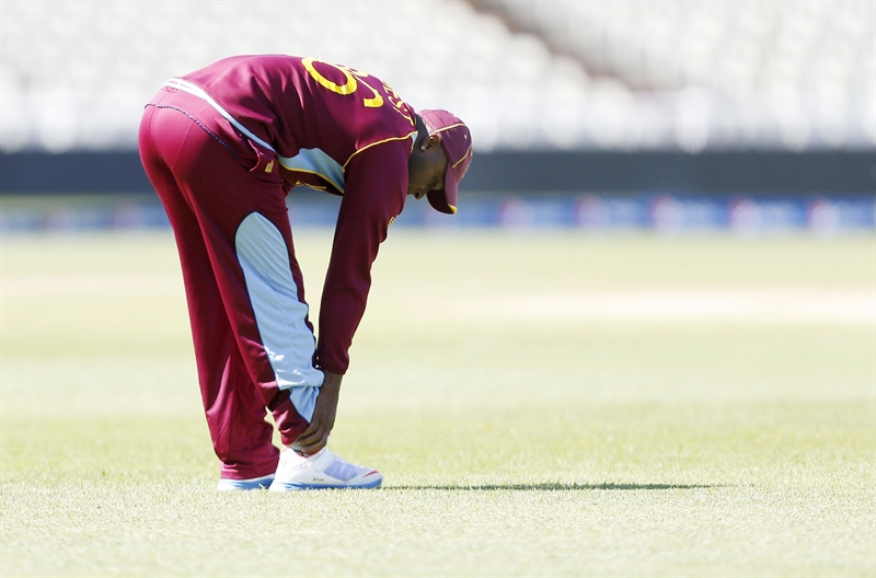
Lateral ankle sprain has a high incidence of occurrence and prevalence amongst the athletic population. In part one of this series, we looked at the natural progression from lateral ankle sprain to chronic ankle instability (CAI) and it was highlighted how after an initial injury, up to 80% of sufferers in high risk sports may end up with long term pain, swelling, dysfunction, and sensations of giving way. The eventual result is CAI(1,2). This suggests that many ankle sprain sufferers will most likely sprain their ankles again, and this may then cascade to late stage CAI.
Although most cases of lateral ankle sprain and subsequent CAI may successfully be treated conservatively through a structured rehabilitation program, this approach may fail, and the athlete is then left with pain, long term dysfunction and instability. In the athletic population, often surgery is advocated in serious first time lateral ankle sprains or if long term CAI develops.
Debate exists in the literature as to whether anatomic repairs are favoured over non-anatomic repairs. The argument for anatomic repair will be discussed here in detail. In the final part of this series next month, the extensive rehabilitation protocol of both anatomic repairs and non-anatomic repairs will be compared and contrasted in detail.
CAI management
ConservativeIt is typical for the sports medicine practitioner to trial a period of conservative management prior to considerations for surgery (see box 1). Only in extreme cases of gross ligament disruption to both the anterior talofibular ligament (ATFL) and calcaneofibular ligament (CFL) will surgery be immediately advocated.
Box 1: Conservative management options prior to considerations for surgery
The characteristic conservative treatment modalities in the management of CAI include(3):- Manual therapy to improve talocrural dorsiflexion. This can include joint mobilisations and manipulations(4-7).
- Manual therapy to the distal fibula(8).
- Soft tissue massage and instrumented-assisted soft tissue (IAST) to the soleus, flexor hallucis longus, tibialis anterior/posterior and peroneals.
- Direct concentric/eccentric strengthening of the lateral ankle stabilisers (peroneals).
- Proximal hip muscle strengthening(9-13).
- Balance and proprioceptive retraining on balance boards, rocker boards, BOSU balls(10-12).
- High-level agility and ankle/knee/hip control activities such as sandpit training and trampoline training.
- Lateral heel supports and orthotics to control rearfoot and midfoot movement.
- Strapping and protective braces to support the ankle(14-17).
In a systematic review conducted by Kerkhoffs et al, it was found that early weight bearing with support following acute ankle sprain improved the overall resolution of symptoms associated with a lateral ankle sprain(17). This early weight bearing progression should incorporate an appropriate supportive boot and this may be followed with taping and soft bracing. Several studies have shown that incorporating early weight bearing and movement improve the return to normal activity, and prevent the development of CAI(17-20).
In part one of this series, we discussed the differences in mechanical instability (gross anatomoical disruption of the ATFL) versus functional instability (balance and proprioceptive dysfunction) of the ankle, and how they can often occur in isolation or in combinations. The reader is therefore directed to issue 163 for a detailed description of the pathogenesis of how an acute ankle sprain leads to CAI.
It is believed that patients with functional instability without demonstrable mechanical instability are more likely to benefit from these non-operative measures mentioned above(21). Surgery is generally reserved for patients with CAI that have failed to improve with a thorough course of conservative management and physical therapy; typically these are the CAI sufferers who exhibit both mechanical and functional instability with episodes of frequent ‘giving way’ of the ankle.
Surgery
If the decision is made to repair the acutely sprained ankle ligaments, or if failed conservative management results in CAI, then the options for surgery can be divided into two broad categories:
- Anatomic repair of the lateral-ligament complex.
- Non-anatomic repair, consisting of an ankle reconstruction using tendon weave procedures (tenodesis).
*Anatomic repairs
The purpose of an anatomic ligament repair is to reconstruct the torn ankle ligaments and capsule to restore the normal ankle anatomy and stability, whilst preserving functional ankle and subtalar joint motion. This can be accomplished with the use of local tissue repair, free tendon graft, or a combination of both. While this type of repair is a technically simpler surgical procedure than non-anatomic reconstruction procedures, its success is dependent on the condition of the injured tissues, and may sometimes require augmentation and/or tenodesis(22,23). In the event of gross ligament disruption with ligament retraction, it may be too difficult for the surgeon to achieve a satisfactory anatomical repair.
The classic anatomic repair was first described by Brostrom in 1966, who reported on the direct late repair of the lateral ankle ligaments in 60 patients with CAI(24). The torn ends of the ATFL were shortened and repaired directly by mid-substance suturing. In 30% of patients the CFL was also repaired. He reported a success rate of 80% with this technique in terms of return to function and patient satisfaction. As a result of this study, this technique is the foundation of the anatomic repair.
Variations of this original Brostrom procedure include imbrication of the mid-substance of the lateral ligaments, and modifications in the suturing of the ligaments through drill holes in the fibula, with or without reinforcement with fibular periosteum. These include the modified versions by Gould, Karlsson, and the ‘split-Evans’ procedure (see figure 1)(25-28).
The functional outcomes of anatomic repairs have been excellent; studies report success rates as high as 87% to 95%(24-29). Outcome variables that are typically measured include range of motion of the ankle, strength of the periarticular ankle muscles, return to preinjury activity level, need for reoperation and late stage post-surgical complications.
Figure 1: Variations of the Brostrom procedure

The benefits of an anatomic repair include the simple surgical approach (and thus a minimisation of tissue disruption and scar tissue formation), the utilisation of local host anatomy while preserving talocrural and subtalar motion, and fewer complications. The most severe complication, although quite rare, is injury to the superficial peroneal or sural nerve.
*Non-anatomic repairs
Non-anatomic reconstructions use tendons (tenodesis) or other types of graft fixation to stabilise the ankle, along with the repair of the native ligaments. While multiple variations have been described, most techniques involve rerouting of the peroneus brevis around the lateral ankle. The more common surgical variations are those developed by Colville, Watson-Jones, Chrisman-Snook and Evans (see figure 2)(30-36).
The major issue with all of these procedures is that the graft does not follow the orientation of the normal ligaments and therefore it does not adequately support anterior talus movement. As a consequence, the patient may experience subtle anterior ‘sliding’ of the talus, whihc may be perceived by the patient as the ankle having a ‘slight give’. The long-term consequence of this talar slide is that excessive shear forces are applied to the talocrural joint. This may accelerate erosion of the articular cartilage on the talus and lead to early onset degeneration and osteophyte formation across the anterior joint margin. Furthermore, these procedures lead to a decrease in subtalar and, to a lesser extent, talocrural motion, and the increased risk of adjacent cutaneous nerve injury(37,38).
Figure 2: Common surgical variations
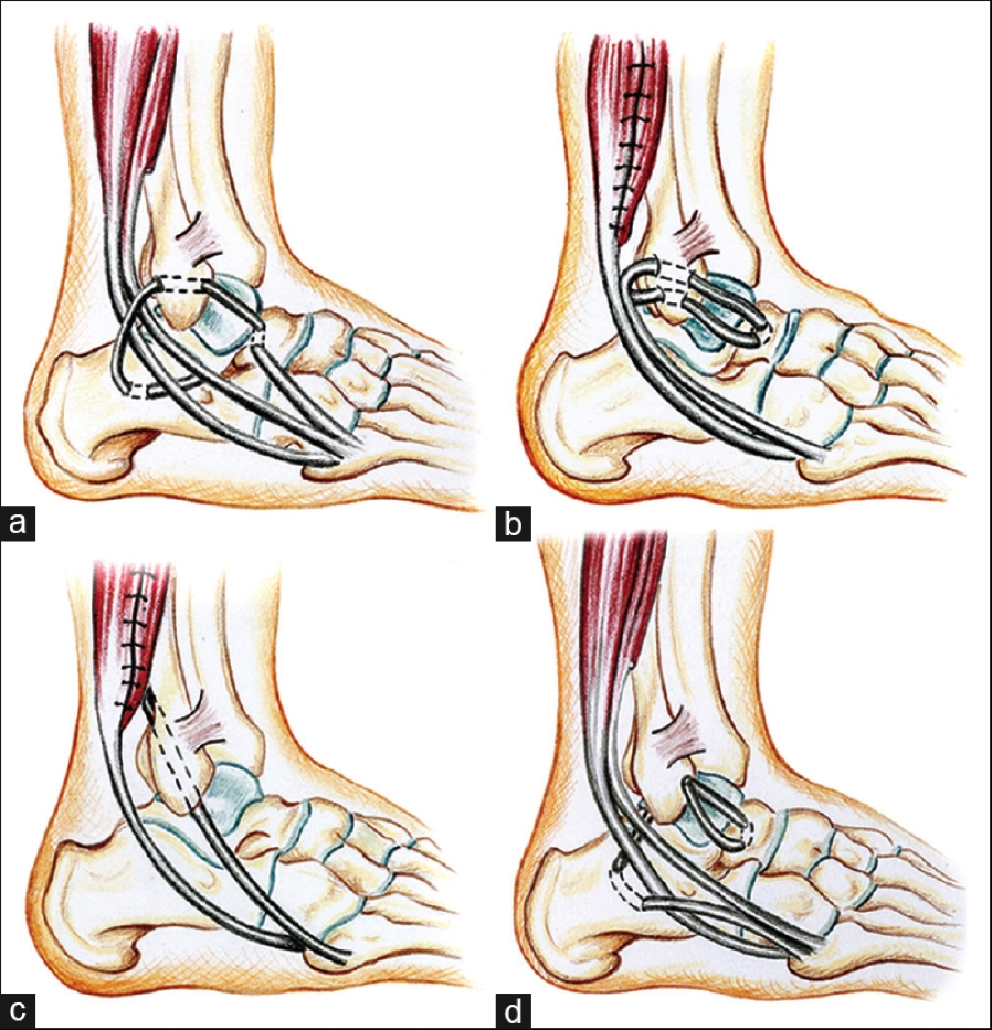
(a) Watson Jones (b) Evans (c) Chrisman Snook (d) Colville (from Al-Mohrej and Al-Kenani 2016 (39)
Comparison studies
Although initial reports on non-anatomic repairs were promising, comparison studies with longer follow-ups generally favour anatomic repair over non-anatomic reconstructions. A host of studies have been published that compare the long term functional outcomes of anatomic and non-anatomic repairs. Below is a summary of some key findings:- In a series of comparison studies, including more than 300 patients, with up to 30 years of follow-up data it was found that long-term, non-anatomic tenodesis led to decreased function, increased pain, limited range of motion, instability, increased need for revision procedures, and greater degrees of osteoarthritis compared to anatomic reconstructions(40, 41).
- In a prospective study that compared the modified Broström with the Chrisman-Snook procedure in 40 patients, it was found that both lead to improvement in more than 80% of patients, although those who underwent a modified Broström procedure had lower rate of complications and better outcomes scores. It was concluded that the modified Broström procedure was superior to the Chrisman-Snook procedure for chronic lateral ankle instability. In addition, a greater proportion of complications occurred with the Chrisman-Snook procedure(42).
- Karlsson et al presented the long-term follow-up of patients with lateral ankle instability treated by the Evans procedure(42). Only fifty percent of patients had satisfactory long-term results. Twelve patients with early satisfactory results had deteriorated at 3-6 years. The absence of normal anatomy resulted in restricted range of motion, reduced long-term stability, and an increased risk of medial degenerative joint disease of the ankle. They found a larger number of reoperations and less satisfactory overall results.
- This was supported by Kaikkonen et al who also found poor results with the Evans procedure(43). Surgical treatment of chronic ankle instability with the Evans procedure restored the mechanical stability of the joint, but all too frequently, the function of the ankle did not return to the pre-injury level. They found that only 35% of their patients achieved an excellent or good result in performance testing. This finding was primarily due to decreased range of motion, swelling of the ankle, and atrophy of the calf.
- Biomechanically, the modified Brostrom procedure was associated with less anterior talar displacement and a decreased talar-tilt angle compared with the Chrisman-Snook procedure(44). The modified Brostrom procedure also roduced a greater mechanical restraint than either the Evans or Chrisman-Snook procedures(44).
Associated surgery
When the surgeon decides with the patient that surgical repair of an ankle with CAI is necessary, often the surgeon will also arthroscope the ankle in attempt to identify and fix intra-articular pathology. Komenda et al arthroscopically examined 54 consecutive patients with lateral ankle instability before ligament stabilisation(45). They noted frequent intra-articular pathology, including a 25% incidence of articular chondral injury. The authors concluded that articular cartilage injuries are common in patients with lateral ankle instability, and can be successfully addressed with ankle arthroscopy in addition to open ligament stabilisation.Advancements in MRI technology may be helpful in identifying associated osteochondral lesions of the talus and peroneal tendon pathology. Its use may be valuable in determining the utility of concomitant arthroscopy, particularly in the presence of mechanical symptoms, chronic ankle pain, or other focal findings on physical examination.
Summary
Although a large number of acute ankle sprain sufferers will respond to conservative treatment, it is possible that long-term CAI may develop. This may then necessitate surgery to stabilise the ankle and allow a return to function and athletic pursuits. On the balance of evidence, it appears that anatomic repairs are favoured over non-anatomic repairs as the normal anatomy of the ankle is preserved and long term outcome studies appear more favourable. In the next issue of SIB, a follow-up article will investigate the extensive rehabilitation protocols of both anatomic repairs and non-anatomic repairs, which will be compared and contrasted in detail.References
- Br J Sports Med. 2001;35:103–108
- J Athl Train. 2002;37(4):376–380
- NYU Hosp Jt Dis 2011; 69: 17-26
- J Am Osteopath Assoc. 2003;103(9):417–421
- Man Ther. 2004;9(2):77–82
- Phys Ther. 2001;81(4):984–994
- J Orthop Sports Phys Ther. 2006;36(7):464–471
- Phys Ther Sport. 2015;16(2):135–139
- Int J Sports Med. 1994;15(6):330–334
- Scand J Med Sci Sports. 1999;9(2):104–109
- J Sport Rehabil. 2010;19(1):98–114
- J Orthop Sports Phys Ther. 1996;23(5): 332–336
- Am J Sports Med. 2006;34(3):464–470
- 2009;373(9663):575–581
- Sports Med. 2011;41(3):185–197
- Foot Ankle Int. 1998;19(10):653–660
- Arch Orthop Trauma Surg. 2001;121(8):462–471
- Am J Sports Med. 1994; 22(1):83–88
- Mil Med. 1994;159(1):20–24
- Sports Med. 1999;27(1):61–71
- Foot Ankle Clin 2006; 11: 531-53
- J Am Acad Orthop Surg 1998; 6: 368-377
- Tech Foot Ankle Surg 2005; 4: 98-103
- Acta Chir Scand. 1966;243:551–565
- Foot Ankle. 1980;1:84–89
- J Bone Joint Surg Am. 1988;70:581–588
- J Bone Joint Surg Br. 1989;71:300–303
- Am J Sports Med. 1989;17:268–273
- Foot Ankle Int. 1999;20:246–252
- Am J Sports Med. 1995;23:210–213
- J Bone Joint Surg Br 76:610–613
- Am J Sports Med. 1996. 24:400–404
- J Bone Joint Surg Am. 1985;67:1–7
- Clin Orthop. 1981;160:201–211
- Am J Sports Med. 1992;20:594–600
- Proc R Soc Med 46:343–344
- Am J Sports Med. 1992;20:594–600
- World J Orthop 6(2): 161-171
- Avicenna Journal of Medicine. 2016. 6(4), 103-108
- Knee Surg Sports Traumatol Arthrosc 2000; 8: 173-179
- Foot Ankle Int 2001; 22: 415-421
- J Bone Joint Surg Br. 1988;70:476–480
- Scand J Med Sci Sports. 1999;9:239–244
- Am J Sports Med. 1994;22:313–317
- Foot Ankle Int. 1999. 20(11), 708-713
Latest Issue
Subscribe Today
Newsletter Sign Up
Subscriber Testimonials
Dr. Alexandra Fandetti-Robin, Back & Body Chiropractic
Elspeth Cowell MSCh DpodM SRCh HCPC reg
William Hunter, Nuffield Health
Subscribe Today
Further Reading
Newsletter Sign Up
Coaches Testimonials
Dr. Alexandra Fandetti-Robin, Back & Body Chiropractic
Elspeth Cowell MSCh DpodM SRCh HCPC reg
William Hunter, Nuffield Health
Subscribe Today
Latest Issue
Be at the leading edge of sports injury management
Our international team of qualified experts (see above) spend hours poring over scores of technical journals and medical papers that even the most interested professionals don't have time to read.
For 17 years, we've helped hard-working physiotherapists and sports professionals like you, overwhelmed by the vast amount of new research, bring science to their treatment. Sports Injury Bulletin is the ideal resource for practitioners too busy to cull through all the monthly journals to find meaningful and applicable studies.
*includes 3 coaching manuals
Get Inspired
All the latest techniques and approaches


Sports Injury Bulletin brings together a worldwide panel of experts – including physiotherapists, doctors, researchers and sports scientists. Together we deliver everything you need to help your clients avoid – or recover as quickly as possible from – injuries.
We strip away the scientific jargon and deliver you easy-to-follow training exercises, nutrition tips, psychological strategies and recovery programmes and exercises in plain English.
