Proximal ITB Enthesopathy
Clinicians frequently miss proximal ITB enthesopathy when diagnosing lateral hip pain. Although uncommon, it can stop athletes in their tracks and warrants greater attention. Evan Schuman uncovers proximal ITB enthesopathy to guide clinical management.
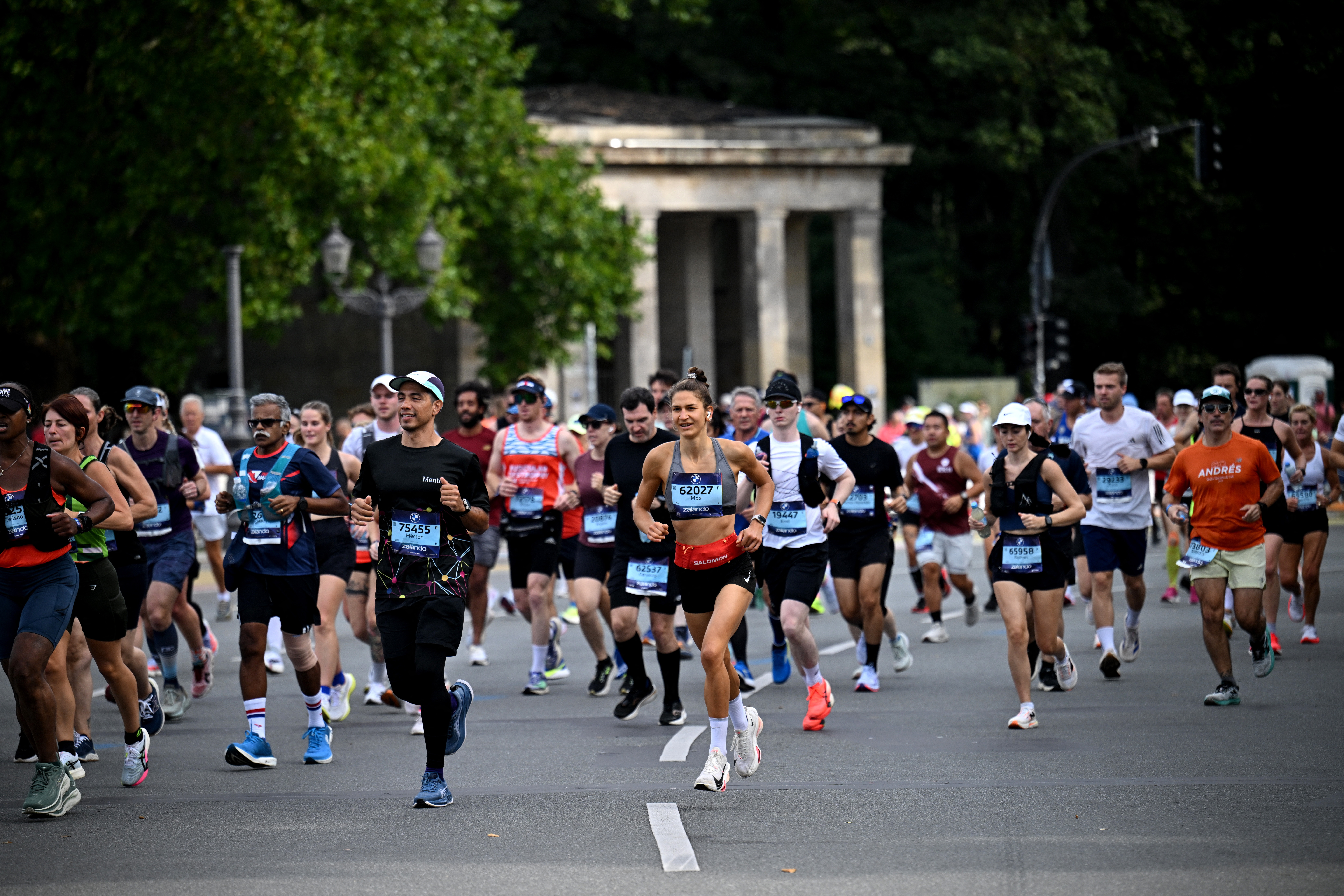
Runners during the marathon REUTERS/Annegret Hilse.
Overuse hip injuries are common in runners due to repetitive microtrauma(1). Common conditions include proximal hamstring and hip flexor tendinopathies, greater trochanteric pain syndrome (GTPS), gluteus tears, and iliac crest stress injuries in adolescent athletes(1,2). Clinicians define enthesopathy as the metabolic, inflammatory, traumatic, or degenerative pathological changes that occur at the enthesis(3). Proximal iliotibial band (ITB) enthesopathy is often misdiagnosed, leading to ineffective treatment. Clinicians should consider this as a potential cause of lateral hip pain.
“Diagnosing proximal ITB enthesopathy can be challenging…”
Anatomy(2,4-6)
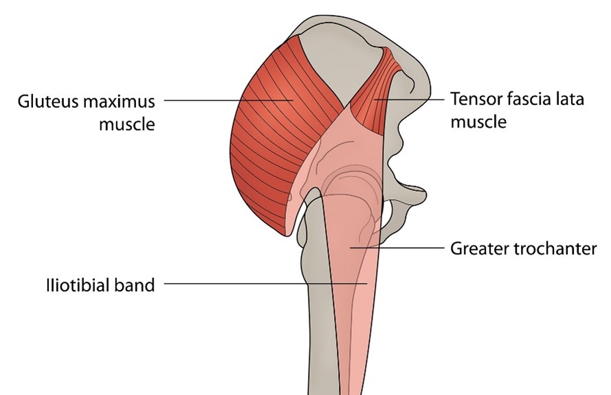
The fascia lata, or deep fascia, is a fibrous structure that envelops the hip and thigh musculature and attaches at multiple sites. The iliac tubercle is a prominent bony landmark on the outer lip of the iliac crest, located five centimeters posterior to the anterior superior iliac spine (ASIS), where the tensor fascia lata (TFL) originates at its anterior portion. Some fibers of the ITB attach to a distinct region of fascial thickening (see figure 1). At the proximal lateral aspect of the hip, the ITB blends with the TFL and gluteal aponeurotic fascia. Distally, the ITB inserts onto Gerdy’s tubercle at the lateral aspect of the tibia.
The enthesis is an area where tendon, ligament, fascia, or joint capsule inserts into bone. Entheses transmit tensile loads from soft tissue to bone and play a vital role by ensuring the efficient transfer of muscle-generated forces to the skeletal attachment, while also dispersing stress away from the enthesis, guiding it from the tendon into the bone. Entheses are classified according to the type of tissue present at the skeletal attachment site, i.e., dense fibrous connective tissue or fibrocartilage. Dense fibrous entheses are commonly attached to metaphyses and diaphyses of long bones, whereas fibrocartilaginous entheses are frequently attached to epiphyses and apophyses. The entheses of the ITB are fibrocartilaginous and are more prone to overuse.
Functions of the ITB
The ITB has tendon-like properties, transferring forces from muscles that attach to it, such as the TFL and gluteus maximus. These muscles fulfil different hip functions. The TFL contributes to flexion, internal rotation, abduction, and pelvic stabilization. The gluteus maximus is a primary hip extensor and assists in external rotation, abduction, and tensioning of the ITB(7).
The ITB assists hip abductors in maintaining a neutral pelvis by preventing lateral pelvic tilt/drop, and it contributes to postural support during single-leg stance and running(2,7). It also stabilizes the knee through muscle tensioning by resisting external adduction forces and applying compressive force to the lateral femur(7). Finally, the ITB attaches the TFL and gluteus maximus to the pelvis, femur, and tibia, giving it tendon-like qualities. These properties provide joint stability and allow the ITB to store and release elastic energy(7).
Straining the ITB
The hip abductors work in two synergies: the trochanteric abductors (gluteus medius and minimus) as primary movers, and the ITB-tensioning muscles (upper gluteus maximus, TFL, vastus lateralis), which stabilize the hip and pelvis by tensioning the iliotibial band(8). During single-leg stance, trochanteric abductors provide about 70% of the abduction force, with ITB-tensioners contributing the remaining 30% via the iliotibial band(9). When the trochanteric abductors are weak, the ITB-tensioners may compensate and increase their stabilizing role. This shift in demand places higher forces on the ITB.
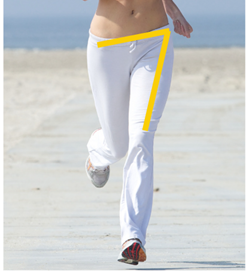
Females demonstrate increased hip adduction torque due to a larger hip width relative to femoral length, along with greater peak hip adduction and internal rotation during both walking and running (see figure 2). These positions increase the strain on the ITB(2). Increased hip adduction strains the ITB because of a greater magnitude of loading, a higher loading rate, or a longer time spent in a strained alignment(10). Weak hip abductors may increase the hip adduction angle(11).
Clinicians must be aware of the biomechanical effects and muscular activation patterns of walking and running on both flat and inclined surfaces. When running on an incline, the hip adduction angle increases, which in turn increases the activity of the gluteus maximus, gluteus medius, and vastus lateralis muscles as the walking and running incline increases, with females showing greater changes than males(12).
In females, hip adduction and internal rotation angles increase as walking and running speed increase. There is also a correlation between increased speed of running and greater activation of gluteus maximus, gluteus medius, and vastus lateralis(12).
A long run may affect running biomechanics through accumulated fatigue. Athletes may exhibit increased knee internal rotation, which can cause a spiraling effect along the longitudinal axis of the ITB, resulting in strain on its proximal attachment(13).
Increasing running cadence reduces mechanical factors linked to ITB strain. A higher cadence decreases peak hip adduction, loading through the hip joint, and vertical loading rate(10,14). Lower cadences may predispose to increased loading of the ITB.
It is essential to recognize that inflammation of the enthesis is a hallmark feature of Spondyloarthritis. Degenerative enthesis changes are associated with non-inflammatory signs due to overuse. There is a growing body of evidence suggesting that metabolic disorders have a significant impact on entheses. Metabolic syndrome is defined as central obesity (waist circumference ≥94 cm in men, ≥80 cm in women) plus any two of the following metabolic abnormalities (see table 1)(15).
Table 1: Metabolic abnormalities
| Criterion | Definition/Threshold |
| Triglycerides | ≥150 mg/dL or treatment for elevated triglycerides |
| HDL Cholesterol | <40 mg/dL (men), <50 mg/dL (women), or treatment for low HDL |
| Blood Pressure | Systolic ≥130 mmHg or Diastolic ≥85 mmHg, or treatment for hypertension |
| Fasting Glucose | ≥100 mg/dL or diagnosed type 2 diabetes, or treatment for hyperglycemia |
The effect of metabolic disorders on enthesopathies:
- Increased BMI places additional load on entheses(15-17).
- Diabetes promotes tendon degeneration and dysfunction through the accumulation of advanced glycation end products(15,16).
- Adipose tissue acts as an endocrine organ, secreting adipokines that contribute to systemic inflammation and vascular dysfunction, further compromising the enthesis(15).
Clinical Presentation(1,2,18,19,20)
- Age 32–67, most often female.
- More common in runners and those with higher BMI.
- Gradual, constant lateral hip pain, often well localized to the iliac tubercle.
- May show a positive Trendelenburg sign.
- ++ tenderness on the iliac crest, just posterior to ASIS.
Assessment(1,2,18,20)
There are no specific tests for the proximal ITB, so clinicians must rely on their knowledge of anatomy, physiology, and clinical reasoning to guide objective testing (see table 2).
Table 2: Objective testing
| Assessment | Rationale |
| Look | Effusion, edema, or abnormal/asymmetrical pelvic alignment |
| Lumbar spine | Clear radicular symptoms |
| Palpation | Local tenderness/pain reproduction |
| Ober’s test | ITB/TFL tightness |
| FABER | Pain provocation |
| Diagonal sit-up | Oblique activation may reproduce symptoms due to the attachment site at the iliac crest |
| Balance - static and dynamic | Postural control deficits |
| Functional tests (single-leg squat/step down) | Observe excessive hip adduction, the Trendelenburg sign |
| Single-leg hopping | Observe lateral pelvic tilt. Clinicians must maintain a high index of suspicion for femoral neck stress fractures if the single-leg hop is painful |
| Running (flat and incline) | Toe in angle/excessive internal hip rotation, hip adduction, knee internal rotation |
| Hip abductor strength | Assess for weakness |
Imaging
Diagnosing proximal ITB enthesopathy can be challenging, as it may mimic other hip pathologies(18). Ultrasound is the first-line imaging modality when pain localizes to the iliac crest, often showing ITB or gluteus medius thickening, disrupted fibrillar architecture, and Doppler hypervascularity(19). An MRI is indicated in chronic or inconclusive cases to rule out stress fractures and other hip disorders, but the field of view (FOV) must include the iliac region(18,19). A small FOV used in hip protocols may miss the proximal ITB(4,18). Clinicians require a larger FOV, including the pelvis and iliac crest, for adequate visualization(4). MRI findings include bright T2 signal at the enthesis, perifascial edema along the ITB, partial-thickness tears near the iliac tubercle, proximal thickening, adjacent soft tissue edema, enthesophytes, and occasionally marrow edema of the iliac tubercle(4).
“++ tenderness on the iliac crest, just posterior to ASIS”
Management
- Pain
A period of relative rest and cryotherapy may help manage inflammation and pain(1). Oral and local anti-inflammatory medications may be beneficial during the acute phase(1). Clinicians may consider shockwave therapy or glyceryl trinitrate patches(6). - Education
Clinicians must educate patients on load management. Patients should also be well-informed about factors that may exacerbate their symptoms. Key considerations include cadence, running mechanics, running surface (flat versus uphill), and postural habits (such as avoiding sitting with crossed legs)(21). - Balance exercises
Single-leg stance exercises may address the challenge of abductor imbalance. It trains coordination between the two abductor synergies (trochanteric and abductor)(22). - Running mechanics
Clinicians must attempt to reduce peak hip adduction during running through interventions such as visual biofeedback and cadence adjustments. Patients may also need to limit their running volume and elevation to avoid fatigue-related compensations at the hip and knee while building the necessary capacity through rehabilitation(21). - Stretching
In gluteal tendinopathy, athletes often avoid gluteal stretches, as these exercises place compressive and tensile loads on the gluteal tendon insertions. The same principles must be applied to protecting the enthesis, and athletes should avoid stretching the lateral hip structures(21). - Strengthening
For proximal ITB enthesopathy, clinicians should emphasize clam, sidestep, unilateral bridge, and quadruped hip extension, which activate the gluteus medius and superior gluteus maximus more than the TFL, thereby strengthening the hip abductors while minimizing TFL dominance(23). The anterior gluteus medius is most active during isometric standing hip abduction and the dip test. The middle segment is active during single-leg bridges, side-lying hip abduction with internal rotation, lateral step-ups, resisted sidestepping, and resisted standing hip abduction. The posterior segment is active during isometric standing hip abduction(24). It is also essential to add an endurance component to these exercises.
Conclusion
Proximal ITB enthesopathy is an uncommon condition with limited literature available. Clinicians face the challenge of making an accurate diagnosis from a presentation they may not have previously encountered. Achieving accuracy requires a thorough understanding of the condition’s presentation, relevant anatomy, physiology, biomechanics, and the factors contributing to ITB strain. Clinicians must then integrate this knowledge into the rehabilitation and management process, ensuring that each aspect of care is guided by sound reasoning and tailored to the needs of their athletes.
References
1. PM R. 2019;11(2):206-209.
2. Skeletal Radiol. 2011;40(12):1553-1556.
3. Semin Arthritis Rheum. 2007;37(2):119-126.
4. Radiographics. 2013;33(5):1437-1452.
5. Muscles Ligaments Tendons J. 2014;4(3):333-342.
6. BJSM.2020 Lateral ‘hip’ pain? Don’t always blame the glutes
7. Sports Med. 2022;52(5):995-1008.
8. Sports Med. 2015;45(8):1107-1119 .
9. Ann Anat. 1993;175(3):203-210.
10. J Sports Sci. 2015;33(7):724-731.
11. J Orthop Sports Phys Ther. 2010;40(2):52-58.
12. Clin Biomech (Bristol). 2008;23(10):1260-1268.
13. Gait Posture. 2007;26(3):407-413.
14. Med Sci Sports Exerc. 2011;43(2):296-302.
15. Reumatol Clin. 2022;18(5):273-278.
16. Clin Rheumatol. 2014;33(10):1517-1522.
17. J Rheumatol. 2020;47(7):968-972.
18. MOJ Orthop Rheumatol 10(1): 00381.
19. JBR-BTR. 2012;95(6):369.
20. Phys Med Rehabil Clin N Am. 2016;27(1):53-77.
21. Sports Med. 2015;45(8):1107-1119.
22. J Orthop Sports Phys Ther. 2015;45(11):910-922.
23. J Orthop Sports Phys Ther. 2013;43(2):54-64.
24. Int J Sports Phys Ther. 2020;15(6):856-881.
Newsletter Sign Up
Subscriber Testimonials
Dr. Alexandra Fandetti-Robin, Back & Body Chiropractic
Elspeth Cowell MSCh DpodM SRCh HCPC reg
William Hunter, Nuffield Health
Newsletter Sign Up
Coaches Testimonials
Dr. Alexandra Fandetti-Robin, Back & Body Chiropractic
Elspeth Cowell MSCh DpodM SRCh HCPC reg
William Hunter, Nuffield Health
Be at the leading edge of sports injury management
Our international team of qualified experts (see above) spend hours poring over scores of technical journals and medical papers that even the most interested professionals don't have time to read.
For 17 years, we've helped hard-working physiotherapists and sports professionals like you, overwhelmed by the vast amount of new research, bring science to their treatment. Sports Injury Bulletin is the ideal resource for practitioners too busy to cull through all the monthly journals to find meaningful and applicable studies.
*includes 3 coaching manuals
Get Inspired
All the latest techniques and approaches
Sports Injury Bulletin brings together a worldwide panel of experts – including physiotherapists, doctors, researchers and sports scientists. Together we deliver everything you need to help your clients avoid – or recover as quickly as possible from – injuries.
We strip away the scientific jargon and deliver you easy-to-follow training exercises, nutrition tips, psychological strategies and recovery programmes and exercises in plain English.
