
 close


 LOGIN
You are viewing 1 of your 1 free articles
Skeletal Muscle: Understanding DOMS
Samantha Nupen reviews how our understanding of delayed onset muscle soreness has evolved from the classic lactic acid and muscle damage theories to newer neural and fascial explanations, and discusses the implications for physiotherapy practice.



Uzbekistan’s Aytjan Khalmakhanov in action during his bout against Japan’s Kyotaro Sogabe. REUTERS/Chalinee Thirasupa.
Few conditions are as familiar to athletes, and yet as poorly understood, as delayed onset muscle soreness (DOMS). For recreational athletes, it is an inconvenience; for elite competitors, it can disrupt training loads, alter biomechanics, and increase injury risk. Yet despite more than a century of research, scientists still debate exactly what causes it.
Delayed onset muscle soreness is characterized by delayed muscle soreness, stiffness, swelling, reduced force-generating capacity, altered biomechanics, and impaired proprioceptive function following exercise(1). Mechanical hyperalgesia, pain on movement and palpation, is the dominant clinical feature(2).
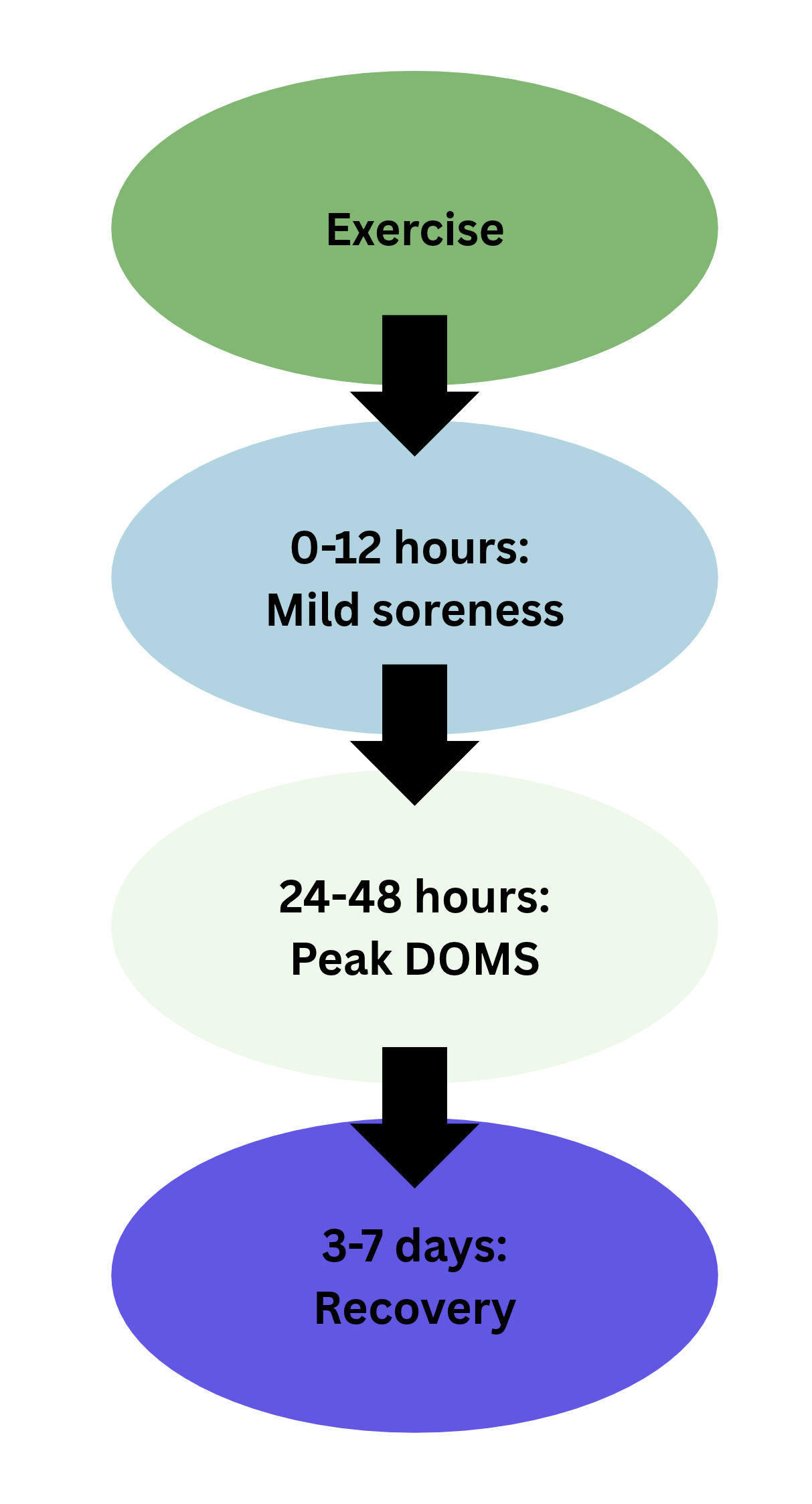
It typically follows unaccustomed or high-intensity eccentric exercise such as downhill running, plyometrics, or heavy resistance training(3,4). Unlike acute exercise-induced pain, DOMS develops after a symptom-free period of 12–24 hours, peaks between 24 and 72 hours, and usually resolves within seven days (see figure 1)(1-3).
The pain-free period makes DOMS somewhat mysterious. Although it is generally self-limiting and does not require medical treatment, the associated loss of strength, altered movement patterns, and reduced proprioception may increase the risk of subsequent injury, particularly in high-performance athletes(1). Thus, preventing and treating DOMS, especially in high-performance sports, is particularly valuable(3).
Early theories attributed DOMS to muscle damage, lactic acid accumulation, muscle spasm, connective tissue injury, and inflammation (see table 1). These hypotheses were gradually replaced as research showed inconsistencies between muscle damage, inflammation, and the timing of soreness(1).
Theodore Hough first described DOMS in 1902 in his classic study in the American Journal of Physiology, titled Ergographic Studies in Muscular Soreness. He proposed a hypothesis of rupture of the muscle fiber (or connective tissue between muscle fibers) as the cause of DOMS because soreness was observed after quick and strong contraction.
"...for elite competitors, it can disrupt training loads, alter biomechanics, and increase injury risk."
Eccentric Exercises
Danish physiologist Erling Asmussen published his foundational observations on DOMS in 1956 in an article titled Observations on Experimental Muscular Soreness in the journal Acta Rheumatologica Scandinavica. He showed that lengthening (negative/eccentric) contractions cause significant delayed soreness one to two days later, whereas shortening (positive/concentric) contractions of equal intensity rarely do. He concluded that the soreness stems from mechanical stress and micro-trauma in the intramuscular connective tissue rather than lactic acid accumulation.
Lactic Acid
Lactic acid was once thought to be the primary cause of DOMS, but this has largely been disproven. Lactate levels return to baseline within an hour after exercise, whereas DOMS develops 12–24 hours later and peaks 24–72 hours afterward. Furthermore, concentric exercise produces far higher lactate levels than eccentric exercise but rarely causes significant DOMS. Likewise, level running generates high lactate concentrations with little soreness, whereas downhill running produces substantial DOMS despite much lower lactate levels. Together, these findings show that lactic acid cannot explain the delayed onset or severity of DOMS(4).
Muscle Damage
Early histological studies demonstrated structural disruption of muscle fibers, including Z-line streaming, sarcomere disorganization, and enzyme leakage such as creatine kinase. These findings formed the basis of the muscle damage theory of DOMS. However, muscle damage does not follow the same time course and similar microscopic muscle damage occurs in pain-free trained athletes. Furthermore, DOMS is observed in cases without muscle damage and anti-inflammatory medication has little effect once DOMS develops.
The Nervous System
Recent research challenges the long-held belief that DOMS is primarily the result of muscle fiber damage. Instead, growing evidence suggests that microscopic injury to sensory nerve endings during eccentric loading may initiate the delayed mechanical hyperalgesia that characterizes DOMS. Animal studies have shown that the time course of nerve growth factor (NGF) production closely mirrors the onset and peak of soreness, offering a plausible explanation for the characteristic 24–48-hour delay that traditional muscle damage theories fail to explain(1).
Further support for this neural mechanism comes from the repeated bout effect–the well-recognized phenomenon whereby DOMS is markedly reduced when the same eccentric exercise is repeated after an appropriate interval. Although the exact mechanism remains uncertain, nervous system adaptations appear to play a significant role. Repeated eccentric exercise reduces both mechanical hyperalgesia and NGF expression, suggesting that neural adaptation may underlie this protective effect(2).
This represents an important shift in thinking, from viewing DOMS primarily as a muscle injury to recognizing it as a complex pain-processing response involving both the neuromuscular and sensory systems.
While neural mechanisms may explain much of the pain associated with DOMS, another emerging theory suggests that the connective tissue surrounding muscle, particularly the fascia, may also play a significant role.
Fascial Involvement
Emerging evidence suggests that the connective tissue surrounding skeletal muscle, particularly the deep fascia, may play a role in DOMS. Rather than an inert covering, fascia is richly innervated with sensory nerve endings and can transmit both mechanical forces and pain. After eccentric exercise, the reduction in pain threshold is greater in fascia than in muscle, suggesting that fascial afferents may contribute significantly to the mechanical hyperalgesia characteristic of DOMS(4).
Fascia is structurally continuous with skeletal muscle through the endomysium, perimysium, and epimysium. This forms an interconnected collagen network that links adjacent muscle fibers and even neighboring muscles. During eccentric contractions, this network experiences substantial tensile loading and may act as a shock absorber, helping to dissipate excessive forces generated within the muscle.
However, when these loads exceed the tissue’s capacity, microscopic damage may occur within the connective tissue itself. As fascia is densely populated with free nerve endings as well as Ruffini and Pacinian corpuscles, it has considerable potential to generate pain and influence proprioception. In the presence of DOMS, the deep fascia becomes more sensitive to noxious stimuli than the underlying muscle, further supporting its role in the development of post-exercise soreness(4).
The emerging fascial model complements the neural theory of DOMS and reinforces the concept that delayed onset muscle soreness is not simply a consequence of muscle fiber damage, but rather a complex neuro-myofascial response to eccentric loading.
Table 1: Historical theory
| Theory | Current understanding |
| Lactic acid | Largely disproven |
| Muscle spasm | Little supporting evidence |
| Muscle fiber damage | Contributes, but does not explain all features |
| Inflammation | Secondary rather than primary |
| Neural sensitization | Strong and growing evidence |
| Fascial involvement | Emerging evidence |
Treatment
Despite decades of research, there is no single intervention capable of preventing or eliminating DOMS. Instead, current evidence suggests that different recovery strategies target different stages of recovery. The goals of treatment should therefore be to reduce pain, restore function, and enable athletes to return to training as quickly and safely as possible(3,5).
Recovery
A wide range of recovery tools has been proposed for athletes. However, evidence supports them to varying degrees. It is vital that clinicians continue to utilize evidence-supported modalities to help demystify the explanations and recovery from DOMS. This ensures that athletes use effective and safe strategies, instead of turning to strategies that may cause further harm to their overall health (see table 2).
Table 2: DOMS recovery strategies
| Recovery strategy | Effect | Level of evidence |
| Cold water immersion (CWI) | One of the best-supported interventions for reducing post-exercise soreness and improving recovery, particularly when used soon after strenuous eccentric exercise. | Good(6) |
| Massage | Reducing the perception of soreness, although improvements in muscle strength and performance are less convincing | Good for perceptual outcomes(6) |
| Foam rolling | Reducing pain and improving mobility, with emerging evidence suggesting it may also enhance fascial sliding. | Moderate(7) |
| Compression therapy | Reducing perceptual soreness. | Emerging evidence(3,6) |
| Heat therapy | Reducing pain and muscle stiffness. | Emerging evidence(3) |
| Functional electrical stimulation | Improving circulation, reducing pain, and supporting muscle function. | Emerging evidence, but optimal treatment protocols remain unclear(8) |
| Collagen supplementation | Collagen peptides (20 g/day for seven days before and two days after eccentric exercise) moderately improved recovery of soreness and jump performance. | Emerging(9) |
| Anti-inflammatories | Anti-inflammatory medication has not consistently reduced soreness or altered the underlying recovery process. | Limited(10) |
"...there is no single intervention capable of preventing or eliminating DOMS."
Prevention
The most effective way to minimize DOMS is through progressive training. Gradually increasing exercise intensity, particularly eccentric loading, allows the musculoskeletal and nervous systems to adapt and takes advantage of the well-established repeated bout effect, whereby repeated exposure to the same exercise substantially reduces subsequent soreness. Performing a small volume of eccentric or maximal isometric exercise several days before an anticipated high-load session may also lessen the severity of DOMS(3).
Clinicians can manage recovery using a multimodal approach rather than relying on a single intervention. Combining cold water immersion, massage, stretching, and functional electrical stimulation at different stages of recovery may provide the greatest benefit by addressing inflammation, pain, and functional restoration simultaneously(8).
Clinical implications
1. Educate athletes that DOMS is a normal response to unfamiliar eccentric loading.
2. Avoid sudden increases in training load.
3. Progress eccentric loading gradually.
4. Don’t equate soreness with tissue damage.
5. Use recovery strategies to optimize training quality rather than eliminate soreness completely.
Conclusion
The clinical understanding of DOMS has evolved considerably. While muscle damage undoubtedly occurs following eccentric exercise, it appears to be only one component of a much more complex neuro-myofascial response. Neural sensitization and the connective tissue network play significant roles. For physiotherapists, this shift reinforces the importance of education, progressive loading and evidence-based recovery strategies rather than relying on treatments aimed solely at reducing inflammation. As research continues to evolve, DOMS is a complex interaction between muscle, connective tissue and the nervous system, with important implications for how clinicians educate, manage and rehabilitate athletes.
References
1. Biomolecules. 2022;12(9):1207.
2. J Physiol Sci. 2016;66(1):43-52.
3. Dtsch Z Sportmed. 2024;75(5):189-194.
4. Int J Mol Sci. 2021;22(17):9482.
5. J Clin Med. 2024;13(7):2006.
6. Front Physiol. 2018;9:403.
7. Front Physiol. 2019;10:376.
8. Front Physiol. 2025;16:1622377.
9. Amino Acids. 2019;51(4):691-704.
10. Rev Bras Med Esporte. 2021;27(6):646-654.

Latest Issue

Subscribe Today
Newsletter Sign Up
Subscriber Testimonials
Dr. Alexandra Fandetti-Robin, Back & Body Chiropractic
Elspeth Cowell MSCh DpodM SRCh HCPC reg
William Hunter, Nuffield Health
Subscribe Today



Newsletter Sign Up



Coaches Testimonials
Dr. Alexandra Fandetti-Robin, Back & Body Chiropractic
Elspeth Cowell MSCh DpodM SRCh HCPC reg
William Hunter, Nuffield Health
Subscribe Today
Latest Issue

Be at the leading edge of sports injury management
Our international team of qualified experts (see above) spend hours poring over scores of technical journals and medical papers that even the most interested professionals don't have time to read.
For 17 years, we've helped hard-working physiotherapists and sports professionals like you, overwhelmed by the vast amount of new research, bring science to their treatment. Sports Injury Bulletin is the ideal resource for practitioners too busy to cull through all the monthly journals to find meaningful and applicable studies.
*includes 3 coaching manuals
Get Inspired
All the latest techniques and approaches


Sports Injury Bulletin brings together a worldwide panel of experts – including physiotherapists, doctors, researchers and sports scientists. Together we deliver everything you need to help your clients avoid – or recover as quickly as possible from – injuries.
We strip away the scientific jargon and deliver you easy-to-follow training exercises, nutrition tips, psychological strategies and recovery programmes and exercises in plain English.
